
There are many patients who in consultation ask us their doubts about the medical follow-up that they will take of their pregnancy achieved thanks to Assisted Reproduction and if said gestational control is different from that which pregnant women undergo spontaneously. For this reason, we have asked Dr. Inés Olmo, gynecologist at the Maternal-Fetal Unit of IVI Valencia, to tell us what the maternal-fetal controls in assisted reproduction pregnancies.

Follow-up after embryo transfer
According to data from National Institute of Statistics (INE), up to 9% of children born in Spain in 2018 were conceived through assisted reproductive techniques and that figure will likely increase in the coming years. For this reason, it is worth paying special attention to monitoring these pregnancies and assisting with their delivery.
The control of the pregnancy of these women is very similar to that of those whose pregnancy has been conceived spontaneously, except for certain peculiarities that I am going to tell you about below.
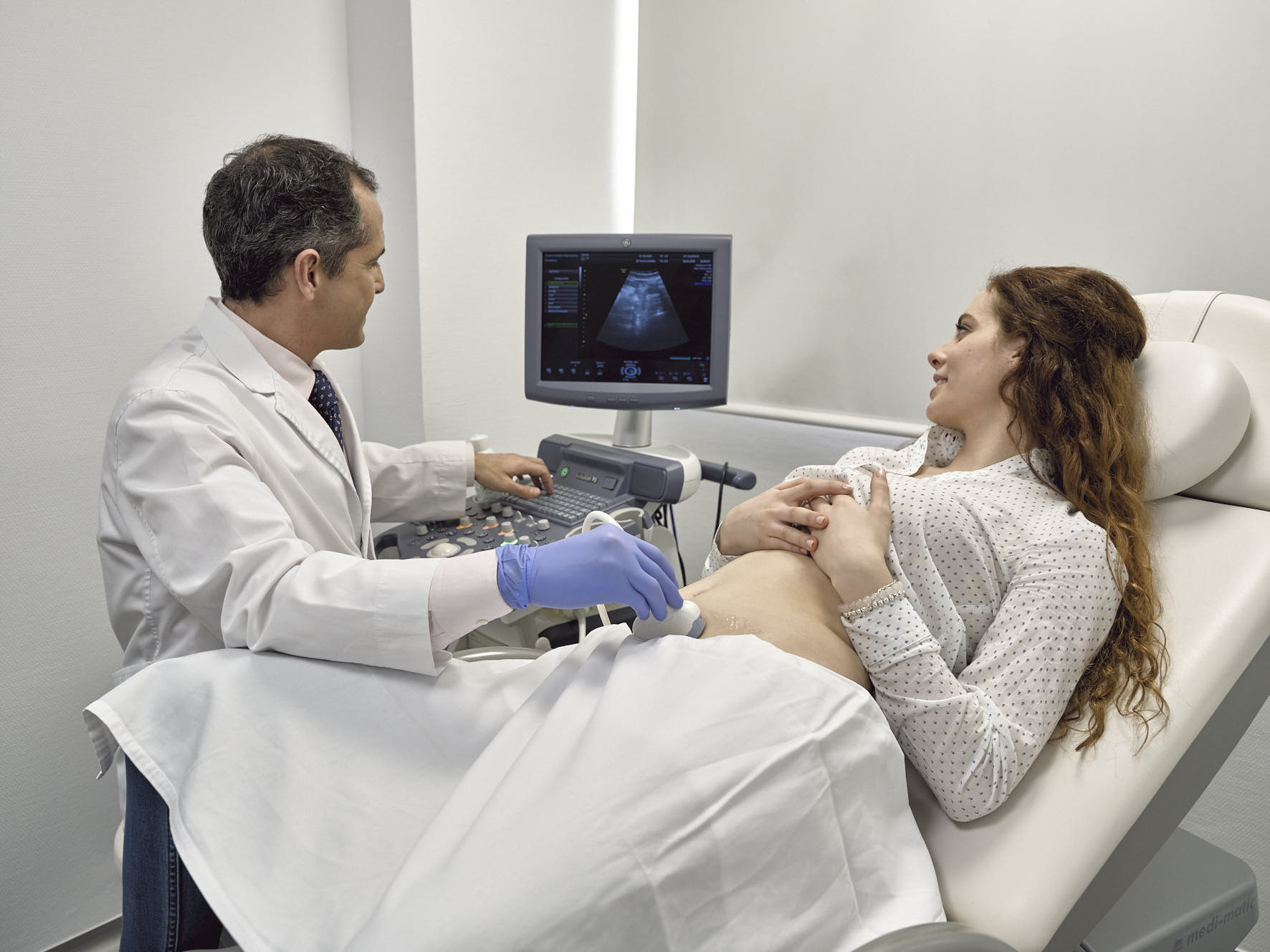
In the first place, in these pregnancies, at 11 days after embryo transfer in In Vitro Fertilization (IVF) or at 14 days in the case of Intrauterine Insemination, we will request an analysis of the β-hCG hormone in the blood (“Pregnancy hormone”). If this is positive, it will indicate pregnancy, and the first ultrasound will be performed between the 5th-7th weeks of gestation, in this way we will confirm the intrauterine location of the gestational sac (ruling out ectopic pregnancy, which is more frequent in this type of pregnancy). We will know if the evolution of the gestation is adequate (early detection of abortions) and we will diagnose if it is a twin pregnancy (they are more frequent in assisted reproduction, although in recent years it is being reduced due to the transfer of a single embryo and the improvement of assisted reproductive techniques).
Gestational controls in the first trimester
Then, as in the rest of pregnancies, a first prenatal consultation is arranged throughout the first trimester, (preferably in week 9-10) that will allow the collection of personal and family medical history of the pregnant woman, performance of a physical examination including taking blood pressure, calculation of BMI, request for a first analysis (includes blood count, TSH, group blood, Rh and presence of irregular antibodies, blood sugar levels, detection of infectious diseases such as hepatitis, HIV, urine culture, cervico-vaginal cytology if necessary). Nutritional advice and advice on physical and occupational activity are provided and specific risks are identified. The first ultrasound will also be requested, already for prenatal diagnosis, which is performed around 12 weeks and is responsible for detecting if the fetus is at risk of chromosomal alterations (Vg sd. Down). To calculate this risk of chromosomal alterations, the same is done in all cases, taking into account the measurement of the nuchal fold obtained by ultrasound, hormonal values collected in maternal blood and the age of the ovum. Here we must bear in mind that in pregnancies after oocyte donation, the age will correspond to that of the donor and not the recipient, in a similar way to when previously vitrified oocytes or embryos are used, the age will correspond to that of the vitrification moment. . It is recommended that in this 12-week ultrasound a Doppler study of the uterine arteries is also performed due to the increased risk of HT in pregnancy (pre-eclampsia) in AR pregnancies.
From here on, successive prenatal consultations are carried out, more or less every 4 weeks, until week 36, and every 2 weeks until week 40, with the pregnancy ending no later than week 41. In them, information on the evolution of pregnancy.
Maternal-fetal follow-up in the second and third trimesters
At second quarter An ultrasound study is carried out between weeks 18 and 22 to detect fetal malformations, which in AR pregnancies follows an identical system to that of the general population, and a second laboratory test (which includes the O’Sullivan test for gestational diabetes screening). Due to the higher rate of multiple gestations and the higher risk of preterm delivery in RA pregnancies, measurement of the uterine cervix by transvaginal ultrasound is advised.
Already in the third trimester An ultrasound scan is indicated around weeks 32 to 36. In the case of AR pregnancies, it is advisable to additionally offer an echocardiographic examination between weeks 28 and 32. The 3rd trimester analysis is requested and the culture is taken against Streptococcus Agalactiae. All pregnant women should be vaccinated against pertussis between weeks 27 to 36. And those Rh negative women will also be administered anti-D immunoglobulin.
As in the rest of pregnancies, all pregnant women will be vaccinated against the influenza virus during the seasonal periods susceptible to contagion.
The most important of gestational control
The termination of pregnancy follows the same criteria as the rest of pregnancies.
Therefore, and finally, we can highlight that:
- Control of a pregnancy achieved by RA is essentially identical that of a spontaneous pregnancy
- Certain peculiarities of pregnancies after AR make it require certain additional actions
Dr. Inés Olmo
Gynecologist of the Maternal-Fetal Unit
IVI Valencia Clinic


