
From IVI we want you to know all the details about what happens inside our laboratories, the place where life is born. For this reason, we have asked Dr. Amparo Mercader, coordinator of the PGT-FIV laboratory at IVI Valencia, to tell you about the process of performing an embryo biopsy.

What is an embryo biopsy?
Embryo biopsy is a technique that is performed in the in vitro fertilization laboratory (IVF), to obtain chromosomal or genetic information from the biopsied embryo. It can be done on day 3 of development or in the blastocyst stage (day 5 or day 6 of development). Depending on the day, the embryos have very different characteristics. When the embryo is on day 3 of development, we can easily count the cells that form it and they are all practically the same.

However, when the embryo is in the blastocyst stage, it is made up of more than 100-150 cells. In this case, what we are looking at is the two parts that make it up. One is the inner cell mass, which is a group of cells that is on the inside that is what will eventually give rise to the baby. The other part is the trophectoderm, which is the outer part and is what will form the placenta.

In which cases is embryo biopsy indicated?
Depending on the day on which the biopsy is performed, the methodology is different, but regardless of this, one must be extremely careful to obtain suitable cells to carry out the analysis and to avoid possible negative effects on the embryo, and this is get experienced. At IVI, we have been doing biopsies since the last century!
The analysis of the samples allows us to select the embryo that is going to be transferred to the uterus in couples with a high risk of transmitting chromosomal abnormalities to the offspring. It is indicated in carriers of karyotype abnormalities, advanced maternal age, recurrent abortion, implantation failure, very severe male factor and previous chromosome disease. Also for genetic abnormalities, so it is indicated in carriers of monogenic diseases. The embryo biopsy is an alternative to the prenatal diagnosis that is carried out after the pregnancy. All of us who work at IVI want our patients to have a live newborn at home, but we also want them to be healthy.
First steps of embryo biopsy
To do it, we need some devices that are already in IVF, such as the laminar flow hood, the incubator and the microscope. But also others that do not have to be in a standard IVF laboratory, such as the laser and the computer. The material we need is practically the same as that used in the IVF laboratory, in addition to specific material to perform the biopsy.
Next, I am going to tell you how the biopsy is performed in the day 3 of embryonic development, that is, when the embryo is in the cell stage. Ideally, the embryo should have between 6 and 8 cells. To be able to analyze a cell we need to be able to access it, so we have to break the zona pellucida (ZP), which is the membrane that surrounds the embryo and protects it from possible adverse conditions outside. There are 3 ways to make the hole: mechanical, chemical or laser. In the IVI we make the hole using a laser, as it is faster, since we want the embryo to be out of the incubator for the shortest possible time. As an image is worth a thousand words, below you have a video in which you can see how it is done.
Once the hole is made, we can obtain the cell using 2 methods: the displacement method or the aspiration method. The first consists of “squeezing” the embryo with a pipette so that the cell comes out of the hole. Aspiration consists of gently aspirating the cell. This is the method we use at IVI and I leave you another video so you can see how we do it.
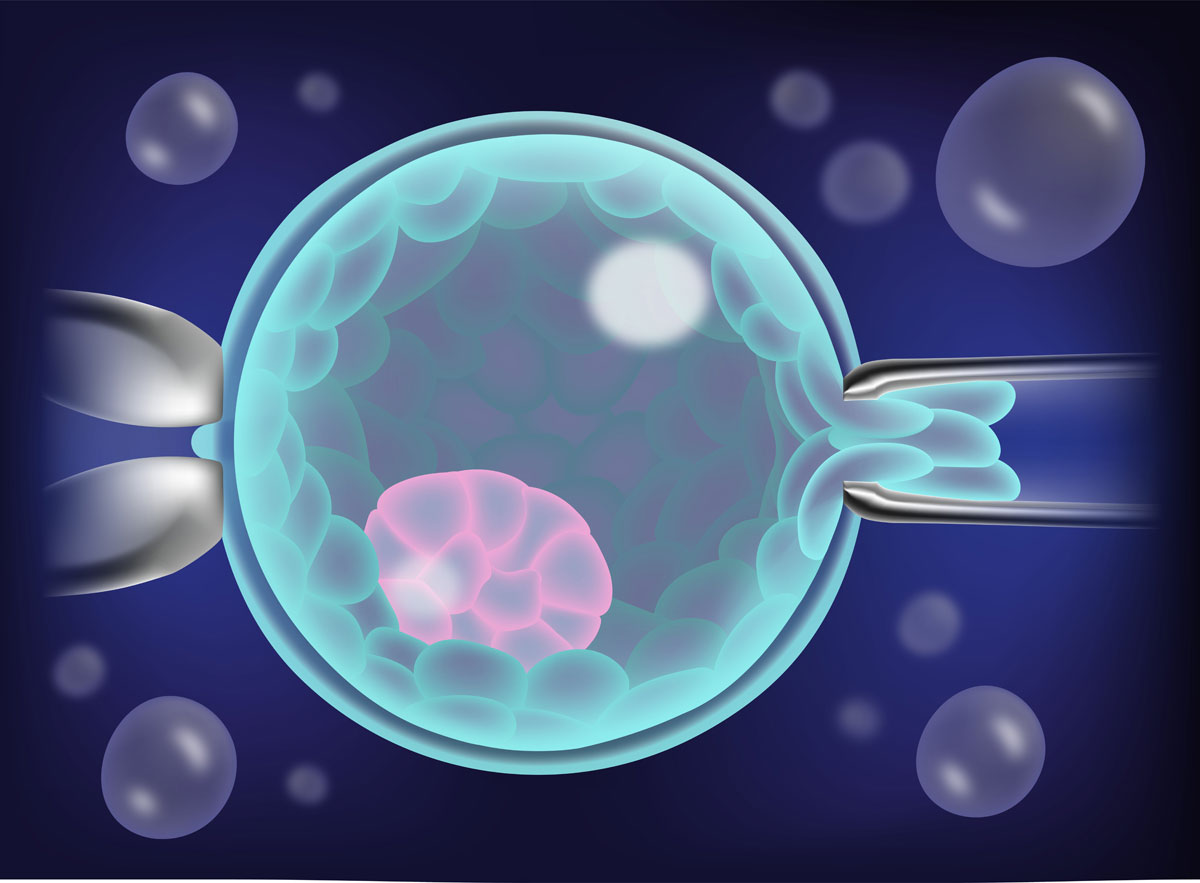
Embryo biopsy in the blastocyst stage
Once the cell is obtained, it will be processed according to the analysis technique that we are going to perform and will be sent to the analysis laboratory. The embryo remains in the IVF laboratory, in the incubator, so that it continues, in the best possible conditions, its development until the blastocyst stage. When we have the results, we inform you and we can transfer the embryo classified as a candidate for transfer. If we have more than one, you already know that the recommended transfer is from a single embryo, the others will vitrify.
The biopsy, as I mentioned at the beginning, can also be performed in the blastocyst stage. This type of biopsy is the one performed in the IVI. I will tell you below how it is. To perform the blastocyst stage biopsy we need the embryo to continue growing until day 5 or 6 of development.
In this type of biopsy we also need to be able to access the cells to send them for analysis. As the blastocyst has many cells, not a single cell is analyzed, but rather a group of cells is taken, about 6, which makes the analysis more robust. To be able to perform this type of biopsy, on day 3 of development, we make a hole in the zona pellucida and let the embryo continue its evolution. With this we achieve that when it reaches the blastocyst, it can leave the zona pellucida, more easily and by itself. This process is called assisted hatching. On day 5 or day 6 of development, see if the blastocyst is leaving the zona pellucida. If so, the cells can be easily accessed and therefore the biopsy can be performed. You look at where the inner cell mass is so as not to touch it, remember that it is the future baby, and several cells are aspirated from the trophectoderm. At this time we can also perform 2 different techniques: a friction movement against the pipette and separate the group of cells or perform laser pulses on the cell junctions and stretch so that the group of cells is separated.
Results of the embryo biopsy
This fragment will be processed, the same as the cell that we obtain when the biopsy is on day 3, depending on the analysis that we are going to perform. In this case, the blastocyst is vitrified pending results. When the analysis laboratory sends us the results, you are informed and a new cycle can be scheduled to carry out the transfer of the blastocyst that has been classified as a candidate for transfer.
The samples that are sent to the analysis laboratory are identified with a clear and unambiguous system that ensures the correspondence with the embryo from which they are derived.
Finally, embryo biopsy is a technique that requires a good deal of practice and experience. But we must not forget that it allows us to transfer embryos with greater potential to give rise to a healthy baby, which is the goal that we all pursue.
I hope I have helped you, with this post, to better understand the embryo biopsy technique.


